Arthroscopy
Arthroscopy was developed for the knee joint for which it is still most frequently used. Now it may be applied to many other joints including the hip & ankles, as well as the shoulder, elbow, wrists & joints. Arthroscopy may be used for diagnosis or for simple or elaborate surgical procedures. Here we will briefly describe its application in the knee joint.
Anaesthesia
Usually general. May also use local or spinal.
Procedure
- A tourniquet is used to provide a bloodless field.
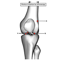
- The knee joint may be accessed at a number of points, the antero-lateral being the most versatile.
- Saline is injected into the joint & through a 1cm skin incision & with the knee flexed, a trochar & arthroscopic cannula are passed. The trochar is removed & a blunt probe is passed through the cannula & into the joint. Remove the blunt probe & pass the arthroscope into the joint.
- Arthroscopes usually comes as a 30° fore-oblique instrument, but a 70° scope may be useful for examining the posterior compartment.
- All compartments are now systematically inspected, & may be opened with the aid of manipulation.
- Through separate portals various instruments may be passed for procedures including biopsy, meniscectomy, patellar shaving, removal of loose bodies, synovectomy, ligament repair etc.
Closure
Squeeze out the saline before withdrawing the instrument. After skin closure apply a firm bandage. Main Postoperative complication: Includes haemarthroses & effusions. Infection occurs in 1-2% & reflex sympathetic dystrophy is unusual but mostly settles.